

Light, confocal and electron (SEM and TEM) microscopes are available at the POLARIS Research Center. Histology and ultramicrotomy labs are also available.
The morphological analyses under service are:
– Preparation and microscopical observation of histological samples from cell culture, organs and small organisms
– Preparation of biological samples for TEM and ultrastructural analysis
– Fluorescent nanoparticles and molecules tracking in cells, organs and small organisms, by different laser confocal microscopy
– Characterization of micro- and nano-particles with SEM and TEM
– Characterization of polymer materials and minerals with TEM e SEM-EDX
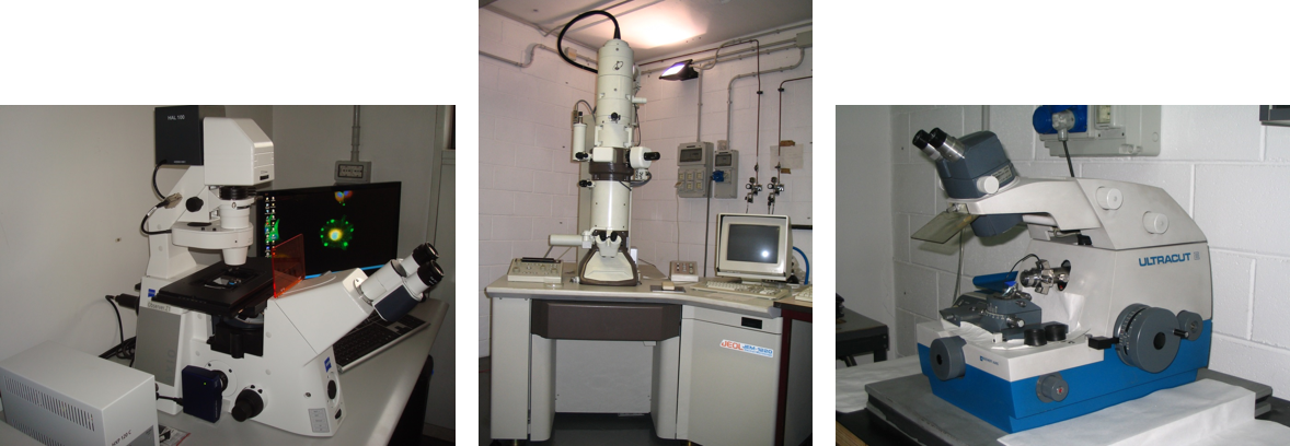
Instrumentations available: Work station Zeiss Axio Observer Z1; confocal microscope Leica SP5- STED; Transmissione electron Microscope Jeol JEM1220; Scanning Electron Microscope TESCAN VEGA 5136XM equipped with a EDX EDAX GENESIS 4000XMS probe; Reichert Ultracut E ultramicrotome.